Fungi


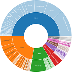
Generally you see only the spore-producing structure (or fruitbody) of a fungus, the rest being well hidden (e.g. in soil, dung or wood). On Nature Map, fungi are divided into categories (the groups and sub-groups shown on the right) based on fruitbody form. Each category name consists of a descriptive phrase and this may be followed:
[ in square brackets ] by a colloquial name
or < in angled brackets > by a technical term
for what you find in that category. Some examples of colloquial terms are mushroom, bolete and polypore – and these are some of the traditional groupings, that have long been used in fungal field guides aimed at the general public.
Why not use just the colloquial terms as category names?
Mushroom is a word that many people are familiar with but it is used in different ways. Mostly it denotes something fleshy with cap, gills and stem (and that will be the meaning in these notes) but some people add the extra condition that a mushroom must be edible (anything else is a toadstool) and some use it virtually as a synonym of fungus. Experience has shown that other traditional terms (e.g. bolete, polypore) are meaningless to people who have never read anything about fungi. The brief explanations in the category names tell users who are unfamiliar with fungi what sorts of fruiting bodies they’ll find in a given category. Note also that some groups of fungi don’t have well-established colloquial names.
If you click on a category you will see an Overview for it. While the category names help, they must necessarily be short and, to get the most benefit, you should read the Overviews. An Overview may (via Hints) offer more detail about the visual or non-visual features that would help identify your sighting or give some Warnings.
The fungi in a given category need not be closely related and the only aim of this categorization is to help you place your sighting. Many users are likely to take photos of fungi incidentally, without necessarily studying the fungi closely, and so would need to rely on just the photograph when trying to place a sighting. For that reason the categories rely on visual features as far as possible to make it easier for those with no knowledge about fungi. The terms in square brackets aim to help those with some knowledge of fungi and even the more knowledgeable users may find more help within the angled brackets.
How to search
If you are unfamiliar with fungi it is best to start at the top of the list on the right and work your way down until you find something that describes your sighting. First are the Cap on a stem groups. In these the top of the fruitbody is sharply differentiated from a supporting stem. In some fungi the fruit body tapers gradually from a broad apex, but with no such sharp differentiation, hence no proper stem (but a pseudo-stem) and such fungi are not included. On past experience, most fungal sightings will be of mushrooms and they are in the first group.
Why do some genera appear in more than one category?
Long ago, fungal classification was closely tied to fruitbody form but later research showed that closely related species need not have the same fruitbody form. Mostly, all species in a genus have the same form of fruitbody but there are exceptions. As an example, look at these two: Schizophyllum amplum and Schizpophylum commune .
This means that in any categorization based only on macroscopic features there will inevitably be cases where all the species of a genus do not fit into the one category. There are a few instances where it is more sensible to deal with slightly dissimilar fruitbody types together and so though your sighting has a fruitbody that should be in category X, a note there will direct you to category Y.
Use of formal taxonomic categories (e.g. genera, families or orders) would avoid the splitting of genera but such categories would not be easy for a non-expert to use since many such categories rely on microscopic features. Also, dissimilar fruitbodies may be grouped together (as Schizophyllum shows) and two species, with fruitbodies of similar form, may fall into different taxonomic categories.
Warnings
The grouping is based on mature fruitbodies and immature fruitbodies may look different.
Many fungi are not identifiable from photos, no matter how good.
With age frutibodies may become tattered, dowdy and unidentifiable from a photo.
More information
For more about fungi in general see: http://www.cpbr.gov.au/fungi/index.html
.....For more about fruitbody types see: http://www.cpbr.gov.au/fungi/types-of-fungi.html
.....For more about macroscopic features see: http://www.cpbr.gov.au/fungi/macroscopic.html


 at Bruce, ACT - 30 Sep 2025 by Hejor1")
 (A polypore) at Bruce, ACT - 30 Sep 2025 by Hejor1")

 at Hawker, ACT - 25 Sep 2025 by CattleDog")

 at Bruce, ACT - 23 Sep 2025 by Lisa.Jok")
 at Ainslie, ACT - 29 Sep 2025 by Hejor1")
 at Hall, ACT - 29 Aug 2025 by Anna123")
 at Chiltern, VIC - 28 Sep 2025 by KylieWaldon")


![Unverified Cap on a stem; gills below cap [mushrooms or mushroom-like] at Kenny, ACT - 24 Sep 2025 by sbittinger](https://api.naturemapr.org/api/sightings/4699999/images/1?width=300&height=300 "Unverified Cap on a stem; gills below cap [mushrooms or mushroom-like] at Kenny, ACT - 24 Sep 2025 by sbittinger")

 at Hall, ACT - 27 Sep 2025 by Anna123")
 at Hall, ACT - 27 Sep 2025 by Anna123")
![Unverified Cap on a stem; gills below cap [mushrooms or mushroom-like] at O'Malley, ACT - 27 Sep 2025 by Mike](https://api.naturemapr.org/api/sightings/4699933/images/1?width=300&height=300 "Unverified Cap on a stem; gills below cap [mushrooms or mushroom-like] at O'Malley, ACT - 27 Sep 2025 by Mike")
 by JimL")

 at Gherang, VIC - 5 Sep 2025 by WendyEM")
 at Gherang, VIC - 5 Sep 2025 by WendyEM")
 at Bonner, ACT - 20 Sep 2025 by Cmperman")
 at Lanitza, NSW - 24 Sep 2025 by Carolina")
![Unverified Pored or somewhat maze-like on underside [bracket polypores] at Symonston, ACT - 24 Sep 2025 by Mike](https://api.naturemapr.org/api/sightings/4699428/images/1?width=300&height=300 "Unverified Pored or somewhat maze-like on underside [bracket polypores] at Symonston, ACT - 24 Sep 2025 by Mike")


![Unverified Cap on a stem; gills below cap [mushrooms or mushroom-like] at Morton Plains, VIC - 14 Jul 2011 by WendyEM](https://api.naturemapr.org/api/sightings/4699183/images/1?width=300&height=300 "Unverified Cap on a stem; gills below cap [mushrooms or mushroom-like] at Morton Plains, VIC - 14 Jul 2011 by WendyEM")


![Unverified Cap on a stem; gills below cap [mushrooms or mushroom-like] at Morton Plains, VIC - 29 Aug 2010 by WendyEM](https://api.naturemapr.org/api/sightings/4699168/images/1?width=300&height=300 "Unverified Cap on a stem; gills below cap [mushrooms or mushroom-like] at Morton Plains, VIC - 29 Aug 2010 by WendyEM")


 at Sherbrooke, VIC - 23 Sep 2025 by Hejor1")


 at Sherbrooke, VIC - 23 Sep 2025 by Hejor1")

 at Sherbrooke, VIC - 23 Sep 2025 by Hejor1")

 at Sherbrooke, VIC - 23 Sep 2025 by Hejor1")
 at Wodonga, VIC - 20 Sep 2025 by KylieWaldon")
 at Kalorama, VIC - 21 Sep 2025 by Hejor1")


 at Mount Dandenong, VIC - 22 Sep 2025 by Hejor1")
 at Mount Dandenong, VIC - 22 Sep 2025 by Hejor1")


![Unverified Cap, gills below, no stem & usually on wood [stemless mushrooms & the like] at Mount Dandenong, VIC - 22 Sep 2025 by Hejor1](https://api.naturemapr.org/api/sightings/4698940/images/1?width=300&height=300 "Unverified Cap, gills below, no stem & usually on wood [stemless mushrooms & the like] at Mount Dandenong, VIC - 22 Sep 2025 by Hejor1")

 (A Coral fungus) at Acton, ACT - 21 Sep 2025 by Clarel")
 at Acton, ACT - 21 Sep 2025 by Clarel")
 at Acton, ACT - 21 Sep 2025 by Clarel")
![Unverified Cap on a stem; gills below cap [mushrooms or mushroom-like] at Acton, ACT - 21 Sep 2025 by Clarel](https://api.naturemapr.org/api/sightings/4698907/images/1?width=300&height=300 "Unverified Cap on a stem; gills below cap [mushrooms or mushroom-like] at Acton, ACT - 21 Sep 2025 by Clarel")

 at The Pilliga, NSW - 21 Sep 2025 by HelenCross")
 at Canyonleigh, NSW - 16 Sep 2025 by blacksheep")
 at Strathnairn, ACT - 21 Sep 2025 by W")
 at Freshwater Creek, VIC - 11 Sep 2025 by WendyEM")
 at Olinda, VIC - 21 Sep 2025 by Hejor1")


 at Olinda, VIC - 21 Sep 2025 by Hejor1")


 at Watson, ACT - 21 Sep 2025 by Lisa.Jok")
![Unverified Cap on a stem; gills below cap [mushrooms or mushroom-like] at Watson, ACT - 21 Sep 2025 by Lisa.Jok](https://api.naturemapr.org/api/sightings/4698663/images/1?width=300&height=300 "Unverified Cap on a stem; gills below cap [mushrooms or mushroom-like] at Watson, ACT - 21 Sep 2025 by Lisa.Jok")
![Unverified Cap on a stem; pores below cap [boletes & stemmed polypores] at Watson, ACT - 21 Sep 2025 by Lisa.Jok](https://api.naturemapr.org/api/sightings/4698662/images/1?width=300&height=300 "Unverified Cap on a stem; pores below cap [boletes & stemmed polypores] at Watson, ACT - 21 Sep 2025 by Lisa.Jok")
 at Watson, ACT - 21 Sep 2025 by Lisa.Jok")


 at Mount Dandenong, VIC - 20 Sep 2025 by Hejor1")
 at Isaacs, ACT - 19 Sep 2025 by Mike")
 at Waurn Ponds, VIC - 14 Sep 2025 by WendyEM")
 at Melbourne, VIC - 19 Sep 2025 by Hejor1")


 at Isaacs, ACT - 18 Sep 2025 by Mike")
 at Franklin, ACT - 13 Mar 2022 by AndyRoo")
 at Franklin, ACT - 13 Mar 2022 by AndyRoo")
![Unverified Cap on a stem; gills below cap [mushrooms or mushroom-like] at Gherang, VIC - 13 Sep 2025 by WendyEM](https://api.naturemapr.org/api/sightings/4697421/images/1?width=300&height=300 "Unverified Cap on a stem; gills below cap [mushrooms or mushroom-like] at Gherang, VIC - 13 Sep 2025 by WendyEM")
 at Kaleen, ACT - 15 Sep 2025 by ConBoekel")
 at Pillar Valley, NSW - 24 Apr 2024 by Topwood")
 at Pillar Valley, NSW - 11 Apr 2024 by Topwood")
 at Tullymorgan, NSW - 2 Apr 2024 by Topwood")
 at Minnie Water, NSW - 24 Oct 2022 by Topwood")


 at Canyonleigh, NSW - 14 Sep 2025 by blacksheep")
 at Hall, ACT - 14 Sep 2025 by Anna123")
Announcements
There are currently no announcements.
Discussion
zz flat polypore - white(ish)
zz - truffle
Truncospora ochroleuca
Some polypore species have very small pores, up to 10 per millimetre. To see such pores you may need to use a magnifying glass or hand lens. A close-up shot with a macro lens would also do the job – but even without that, if you enlarge a photo you my see the pores, depending on the quality of the photo. Here (https://www.neotropicalfungi.com/wp-content/uploads/2024/04/ANGE1885-Rigidoporus-microporus_risultato.jpg) is a photo of Rigidoporus microporus. In a description of this species you’d read that there are 6-9 pores per millimetre. One specimen has been turned to show the orange-brown underside and there are no obvious pores. If I enlarge this photo I still don’t see any pores but soon get into a pixelated image. Perhaps the original photo would show the pores – or perhaps the camera was not close enough to allow pores to show in any magnification of the original photo.
Sometimes a tiny-pored underside may show a slightly fuzzy look, since a smooth surface reflects light differently to a surface with numerous tiny pores. Your photo of the underside is a little blurry. If the edges had been sharp and the rest of the undersurface had been slightly fuzzy (but not obviously out of focus) I’d have suspected a pored surface.
zz flat polypore - white(ish)
zz flat polypore - white(ish)
Significant sightings
- Humidicutis arcohastata at QPRC LGA
- Fomitopsis maculatissima at QPRC LGA
- Lactarius turpis at QPRC LGA
- Boletellus emodensis at Glen Allen, NSW
- Kgaria sp. (genus) at Conder, ACT
- Arachnocrea scabrida at Bruce, ACT
- Myriostoma australianum at Cook, ACT
- Resupinatus sp. at Wattamolla, NSW
- Calocera sp. at Wattamolla, NSW
- Geastrum tenuipes at Broughton Vale, NSW
Top contributors
- trevorpreston 1.2K
- Teresa 825
- TimL 576
- KenT 470
- LisaH 457
- AlisonMilton 421
- Hejor1 343
- Csteele4 284
- Paul4K 221
- Mike 218
Top moderators
- Heino1 4.3K
- Heinol 1.7K
- Teresa 986
- Heino 737
- Pam 581
- Csteele4 563
- MichaelMulvaney 557
- KenT 445
- JTran 151
- CanberraFungiGroup 110